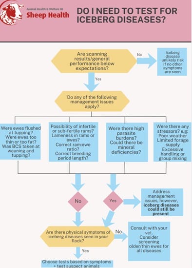

There are 5 iceberg diseases of concern to sheep in the UK and Ireland. These are diseases that are difficult to detect and diagnose, as sheep that show clinical signs are usually just the ‘tip of the iceberg’. This means there can be a large percentage of the flock infected that aren’t showing obvious clinical signs, therefore, usually by the time disease is detected a significant proportion of the flock will be already infected.
These diseases tend to have a long incubation period before showing signs, mainly affecting older ewes, but can subtly be reducing production long before this. There are no treatments for them and they will usually result in fatalities.
UK and Ireland prevalence for iceberg diseases is not well known as they are under diagnosed, but it is likely that many have a strong foothold across many flocks. It is important to understand the significance and recognise the signs of these diseases, as once they are in your flock they can have devastating consequences.
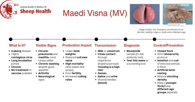
1. Maedi Visna (MV)
Summary
Maedi Visna is a virus that causes lung disease and chronic wasting in sheep. Animals can become infected at any point in their lifetime. The prevalence of this virus is thought to have doubled in GB over the past 10 years. Characteristic of most iceberg diseases, the virus is slow to show symptoms after infection and by the time symptoms do begin to show, a large percentage of the flock is likely to be infected. There is no treatment for this disease, the best method of prevention is to keep it out of your flock.
Signs You May See In Your Flock
Clinical signs are mainly seen in older sheep, and can be seen as:
- Chronic pneumonia
- Dry cough
- Wasting/weight loss (despite a good appetite)
- Arthritis
- Chronic mastitis- hard and lumpy udders, that don’t appear painful
- Neurological signs: muscle tremors and/or incoordination (less common)
- Toe dragging/hind leg paralysis
Some symptoms are less obvious and are affecting productivity:
- Lower lamb weights- lambs may be noticeably hungry
- Reduced milk production
- Premature culling of ewes
- Poor fertility
Economic Losses
- Higher mortality rates (lambs and ewes)
- Increased number of replacements required
- Lower value of cull ewes
How Infection Spreads
- Lambs ingesting infected milk
- Close contact, spread via respiratory droplets or aerosols- housing and at feeding troughs are high risk areas
- Semen of infected rams
- Saliva and urine
Disease Diagnosis
Blood or milk samples can be tested to assess if animals are infected by testing for antibodies to the virus. ELISA tests have been found to be more sensitive than older methods such as AGID. However, there can be a significant period between initial infection and the production of antibodies. Therefore, these tests are not good at detecting early infections.
Testing thin ewes is often used as a screening test, it’s also recommended that all rams are blood tested.
A post-mortem of thin cull ewes or any deaths with similar symptoms to MV can also be valuable. In an infected sheep this will show lungs that are heavy, rubbery, do not collapse and are a mottled, grey colour.
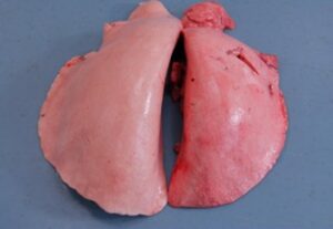
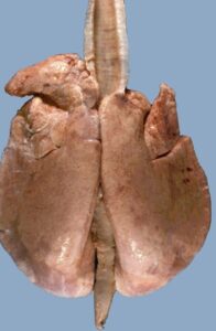
(Image credit: Hal Thompson and Richard Irvine- a pair of healthy lungs (on left) compared to lungs infected with Maedi Visna (on right).
Prevention
Keeping MV out of your flock through excellent biosecurity is the best line of defence, once it has established within your flock, it will spread quickly and is difficult to control.
Biosecurity
- Ensure fencing is in good order- avoiding mixing between neighbouring flocks
- Thick hedges or double fencing to avoid nose-to-nose contact
- Test any rams entering the flock
- Keep a closed flock
Control
There are no treatments or vaccines available for MV. However, there are management options that can be considered to control infection.
Artificial Rearing
Artificial rearing has been found to be an effective method of reducing spread of MV from ewe to lamb. This involves removing lambs from ewes immediately at birth, and feeding them either artificial colostrum or colostrum from a known MV free ewe. These lambs must be reared separately to the rest of the flock and all equipment kept separate and well disinfected. This approach involves a lot of labour and can be costly, however for those with high value stock it may be an effective way to preserve desirable genetic profiles.
Embryo Transfer
There is some evidence that has shown embryo transfer could be another effective way to preserve high value genetics, by taking embryos from MV positive ewes and transferring them to a MV negative ewe.
To minimise the risk of virus transfer, embryos must be collected, processed and transferred in strict accordance with International Embryo Technology Society (IETS) protocols. These internationally recognised standards are designed to reduce the risk of transmitting infectious agents during embryo transfer.
Under IETS guidelines, embryos must be:
- Washed a minimum of 10 times immediately after collection in a suitable sterile medium (for example, phosphate-buffered saline, PBS), with each wash using a fresh pipette and clean medium
- Inspected microscopically between washes to ensure the zona pellucida remains intact and free from debris
- Treated with trypsin following washing, which is used to remove or inactivate any virus particles that may be attached to the outer surface (zona pellucida) of the embryo
Only embryos that meet IETS quality and handling standards should be transferred into MV-negative recipient ewes, and all procedures must be carried out by appropriately trained and accredited personnel using dedicated, disinfected equipment. This is a very costly and labour intensive process but could prove valuable to pedigree flocks where genetic preservation is a priority.
Hard Cull
Some flocks may choose to cull all positive animals within their flock in order to stop disease spread. However tests can miss infected animals and so repeated testing combined with management changes will be necessary to maximise the likelihood of effective long term control.
Split Flocks
For flocks that discover they have a high proportion of positives, they may choose to keep a ‘positive’ and ‘negative’ flock, gradually culling the positives. Management and biosecurity must be very strict in these instances, as the virus is highly contagious.
Goats can also be a possible source of infection, through the closely related virus ‘CAE’, try to keep goats and sheep separated where possible.
Keeping a Younger Flock
By keeping a younger flock and consistently culling older ewes, the chance of MV having a negative impact on production is limited
Reduce Stocking Density/Outdoor Systems
By giving sheep more space, particularly at housing, this will limit the rate of disease spread. While this approach is not suitable for all flocks, research shows that sheep kept outdoors year-round have a much lower risk of MV spreading. In some cases, the disease may even become self-limiting due to the lack of close, prolonged contact between sheep.
2. Ovine Johne’s Disease (OJD)
Summary
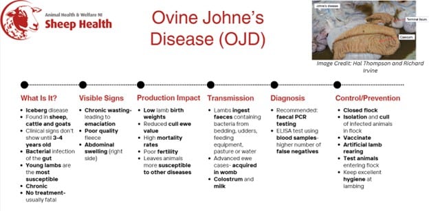
Johne’s disease is caused by a bacteria called Mycobacterium avium subspecies paratuberculosis (MAP). It is found in many animals including sheep, goats, cattle, deer and camelids. It is known as a ‘chronic wasting disease.’ The bacteria causes the intestinal lining to thicken, severely impairing nutrient and water absorption. The disease has a long incubation period, and clinical signs usually start to appear in middle aged sheep (3-4 years old). Lambs are at the highest risk of picking up Johne’s disease. Adult sheep are more resistant to becoming infected. It is a widely undiagnosed infection in sheep and is believed to be very widespread in UK sheep flocks. Mortality rates can be variable. In many cases infection will lead to excessive culling rates. There are no treatment options for Johne’s disease. Any animal showing clinical signs will invariably die from the infection or from another infection the animal has been pre-disposed to.
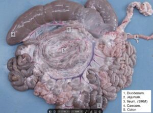
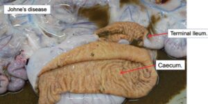
(Image credit: Hal Thompson and Richard Irvine- the first image shows a healthy gut vs. one that has been infected with OJD. Notice the yellow pigmentation of the caecum and terminal ileum, which is characteristic of OJD. It has also caused a thickening of the lining of the caecum and ileum, which is due to an inflammatory reaction, and results in poor absorption of nutrients).
Signs You May See In Your Flock
Infected sheep may appear bright with a normal appetite, however symptoms that will be seen in middle aged ewes include:
- Chronic weight loss (leading to emaciation)
- Poor quality and ‘open’ fleeces
- Bottle jaw
- Abdominal swelling (due to fluid) especially on the lower right side
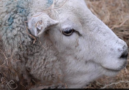
(Image credit: Hal Thompson and Richard Irvine- Bottle jaw)
Economic Losses
- Lambs born to ewes in the advanced/terminal stages of Johne’s have very low birthweights
- Premature culling of ewes
- Lower value of cull ewes
- Animals can be more susceptible to other conditions e.g. parasite burdens
- Poor fertility
How Infection Spreads
- Lambs ingesting faeces through contaminated bedding, udders, feeding equipment, pasture and water
- Can be acquired in the womb. This is more likely in ewes showing clinical signs of infection.
- The bacteria are very resilient and can survive for months on pasture.
- Transmission can happen via colostrum/milk, through both direct spread of MAP into milk as well as faecal contamination of teats and udders.
Disease Diagnosis
- Post-mortem- this is most useful on any animals that were showing clinical signs. Sub- clinically or pre-clinically infected animals are less likely to show pathological changes detectable at post-mortem.
- Faecal test- either a culture or PCR test can be used. Culture is very slow, particularly for sheep strains of MAP (taking up to five months) and isn’t routinely used. PCR test is the preferred method, detecting the DNA of MAP bacteria in It has a much quicker turnaround (7-10 days) and a lower cost than the culture test. It can also be applied on pooled samples and so can be an effective flock screening tool. However the sensitivity of PCR is poor, especially when applied to pre-clinically infected animals, and so it can miss infected animals.
- Blood test- an ELISA test can be used that detects antibodies to the MAP organism. However, like PCR, this test lacks sensitivity (meaning more false-negatives) particularly in pre-clinically infected animals. However, it can be a useful test if applied to carefully selected animals.
It is recommended that you always consult your private vet on what tests may be most appropriate to be used if Johne’s is suspected in your flock.
Prevention
- Keep a closed flock
- Test any animals entering the flock
- Avoid co-grazing with cattle or goats, as it can spread between species
- Don’t graze lambs on pastures recently applied with manure/slurry from suspect animals or a farm of unknown Johne’s status
Control
Vaccination
There is a vaccine licensed for sheep against Johne’s. It’s recommended to vaccinate lambs between 4-16 weeks of age. The vaccine delays shedding of bacteria from infected sheep, reduces the amount shedded and can decrease mortality rates. Vaccination should be discussed with your private vet to see if it is suitable for your situation. Note that there are no blood tests that can differentiate a vaccinated from an infected animal and so care needs to be taken on the implications of vaccination on the ability to detect infection.
Manure management
Once you have established the infected sheep in your flock, you can keep young lambs born to negative testing ewes away from the manure of infected sheep, by keeping good hygiene at lambing. Avoid grazing young lambs on pasture that may be contaminated with MAP for as long as possible, and ensure water troughs are kept clear from manure contamination.
Artificial rearing of lambs
Artificial calf rearing has been successfully used in the dairy industry to reduce Johne’s disease, and the same principles could be used in the sheep industry. If you know there is a problem with Johne’s in your flock, removing lambs at birth and feeding them artificial colostrum or known Johne’s free colostrum can reduce the chances of the lamb picking up Johne’s Disease at the most susceptible age.
Test-and-cull
Removing infected animals from the flock is a crucial element of control but should always be combined with other management strategies.
Breeding Replacements
Infection tends to pass from dams to offspring, through in utero transmission, consumption of colostrum/milk and exposure to dam faeces. The offspring of infected ewes have a much greater likelihood of acquiring infection compared to other lambs. Therefore, it is advisable not to retain the offspring of infected ewes for future breeding, but instead to fatten for slaughter.
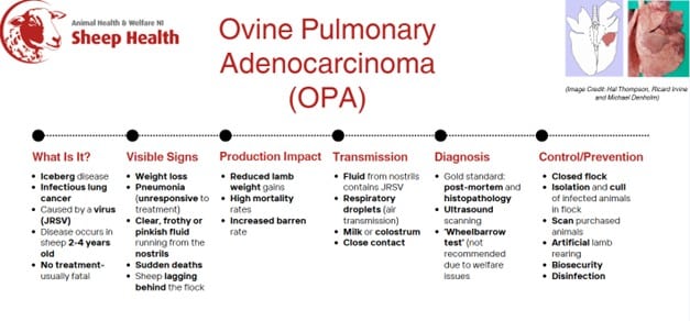
3. Ovine Pulmonary Adenocarcinoma (OPA)
Summary
Ovine Pulmonary Adenocarcinoma (OPA) is an infectious lung cancer in sheep, causing the development of fatal lung tumours. It’s caused by jaagsiekte sheep retrovirus (JSRV). It causes progressive respiratory distress and weight loss, ultimately ending in death. Clinical signs don’t usually become evident until sheep are 2-4 years old, although disease may occur quicker in lambs that are progeny of infected dams (8-12 months old).
Maedi Visna can coexist with OPA in an animal, therefore if clinical signs similar to the two diseases are shown, it may be worth testing for both.
Signs You May See in Your Flock
Usually once clinical signs show, the affected sheep die within days to months.
- Progressive weight loss (with normal appetite)
- Pneumonia (doesn’t respond to treatment, secondary bacterial infections of the lung can also be common)
- Sudden deaths (often due to pasteurella pneumonia- sheep may have been in good body condition)
- Sheep lagging behind flock due to breathing difficulty
- Advanced cases- significant amount of fluid running out of animal’s nostrils when head is lowered (seen in two thirds of cases)
- Cases may peak in adverse weather conditions
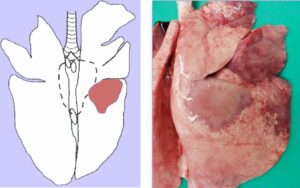
(Image credit: Hal Thompson, Richard Irvine and Michael Denholm- OPA forms as a tumour in the lungs)
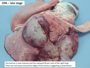
(Image credit: Hal Thompson and Richard Irvine- the progression of the OPA tumour)
Economic Losses
- Increased barren rate
- Reduced lamb weight gains
- High mortality rates
How Infection Spreads
The JRSV virus is found in the fluid from the lungs of infected sheep. Therefore transmission can occur in these cases:
- Through the air via respiratory droplets
- Close contact- housing and trough feeding
- Via milk/colostrum of infected sheep
Disease Diagnosis
The ‘wheelbarrow test’ has been a common diagnosis. It involves lifting the sheep’s hindquarters to lower the head. If a volume of frothy, clear or pinkish fluid flows from the nostrils this is a positive test for OPA. However, this method can cause significant distress to the animal, and an absence of fluid does not rule out OPA.
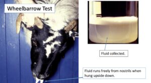
(Image credit: Hal Thompson and Richard Irvine)
There are no commercially available laboratory tests for OPA, as there is a lack of an immune response to JRSV in sheep, meaning no detectable antibodies are produced. The only option for live animals is ultrasound examination of the lungs. Using ultrasound can detect tumours over 1cm in size in the lungs- therefore it may not pick up very early stages of infection and other pathological changes can sometimes be mis-attributed to OPA. Therefore it is crucial that it is carried out by a skilled and experienced operator.
Post-mortem followed by histopathology is the most accurate method of diagnosis.
Prevention
- Keep a closed flock
- Ultrasound scan any purchased animals
- Buy from a trusted source
- Good biosecurity (double-fencing, thick hedges)
Control
Reduce stocking density
By reducing stocking density, this should reduce the speed of spread of the virus, as sheep will not be in as close contact with each other. When feeding, leave plenty of space that sheep do not have to fight against each other, consider using a snacker for meal feeding.
Outdoor lambing
This will not be suitable for every flock, but if possible within an infected flock this will also reduce the speed that the virus spreads, as during housing there is a much higher risk of spread.
Keep sheep in groups according to age
Young animals are the most susceptible and there is a higher transmission risk in older animals that may have advanced OPA lesions. Therefore by keeping the two groups separate this may allow unaffected replacements to be kept.
Artificial rearing of lambs
Artificial rearing could be an effective method of reducing spread of OPA from ewe to lamb. This involves removing lambs from ewes immediately at birth, and feeding them either artificial colostrum or colostrum from a known OPA free ewe- although this can be difficult to determine. These lambs must be reared separately to the rest of the flock and all equipment kept separate and well disinfected.
This approach involves a lot of labour and can be costly, however for those with high value stock it may be an effective way to preserve desirable genetic profiles.
Scan-and-cull
Ensure any ewes scanned positive for OPA are culled.
Cull ewes in poor body condition
Where other possibilities for poor body condition are eliminated (e.g. parasite burden), culling any ewes in poor body condition, have respiratory issues or are seen to be lagging behind the flock may be effective in control, due to the costliness of ultrasound.
Disinfect buildings and equipment that may have been used with infected animals
The fluid that runs from infected sheep’s nostrils contains JRSV and therefore the environment where infected sheep have been is a key area where infection of other animals can occur.
4. Caseous Lymphadenitis (CLA)
Summary
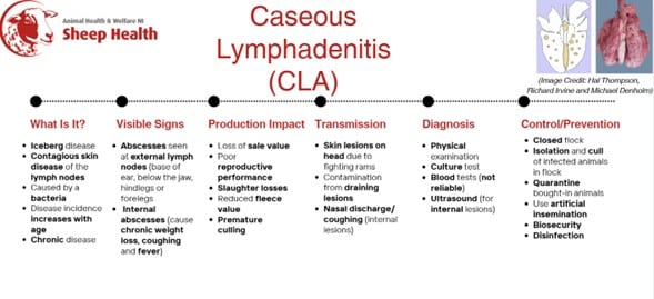
Caseous Lymphadenitis (CLA) is a chronic contagious skin disease that can be seen in sheep and goats. It is caused by a bacteria (Corynebacterium pseudotuberculosis). It’s usually characterized by the formation of abscesses appearing on superficial lymph nodes but can also be found in internal lymph nodes. It’s endemic in Northern Ireland.
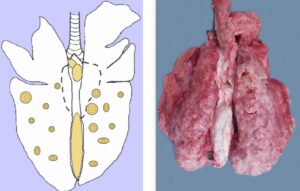
(Image credit: Hal Thompson, Richard Irvine and Michael Denholm- CLA forms abscesses across lymph nodes in the lungs)
Signs You May See in Your Flock
In the UK abscesses are mainly seen in external lymph nodes:
- At the base of the ear
- Below the jaw
- At the hindleg or foreleg
However, infection can occur within lymph nodes of the chest and internal organs (lungs, spleen, kidneys and liver). This can cause:
- Chronic weight loss
- Coughing
- Nasal discharge
- Fever
Development of clinical signs mainly increases with age.
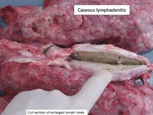
(Image credit: Hal Thompson and Richard Irvine)
Economic Losses
- Reduced sale value- due to unsightliness of abscesses
- Internal lesions may cause poor reproductive performance
- Slaughter losses- carcass may be condemned due to internal lesions
- Reduced fleece value (if contaminated with abscess contents)
- Premature culling
How Infection Spreads
- Rams fighting, causing skin lesions on the head where the bacteria can then enter
- Contamination of surfaces from draining lesions
- Can be transmitted via nasal discharge or coughing by animals with internal lesions
Disease Diagnosis
Physical examination of external abscesses (on lymph nodes), followed by a culture test for the presence of the causative bacteria- taken from material of an abscess. This is thought to be the most effective method of confirmation, however, it cannot be used to detect subclinical cases.
Blood tests can also be used for detecting CLA at flock level, but vary in sensitivity and specificity, producing some false negatives. These are recommended for screening of a flock, rather than identifying individual cases.
Ultrasound can be used for internal lesions and therefore could be used in conjunction with scanning for OPA.
Prevention
- Keep a closed flock
- Quarantine bought-in animals for at least two months and observe for lesions developing
- Use artificial insemination rather than buying in rams
Control
Surgically drain major abscesses
This will provide some relief for the animal and may reduce potential disease spread, however in most cases lesions will return so achieving a cure is very unlikely.
Isolate and cull infected animals
Immediately isolate any animals with suspicious lesions and get them examined by your private vet. If they are confirmed CLA positive, cull these animals to prevent further spread within the flock.
Thorough disinfection of all areas infected animals may have been
Pus from the lesions will infect other animals, therefore thorough cleaning and disinfection is essential.
5. Border Disease (BD)
Summary
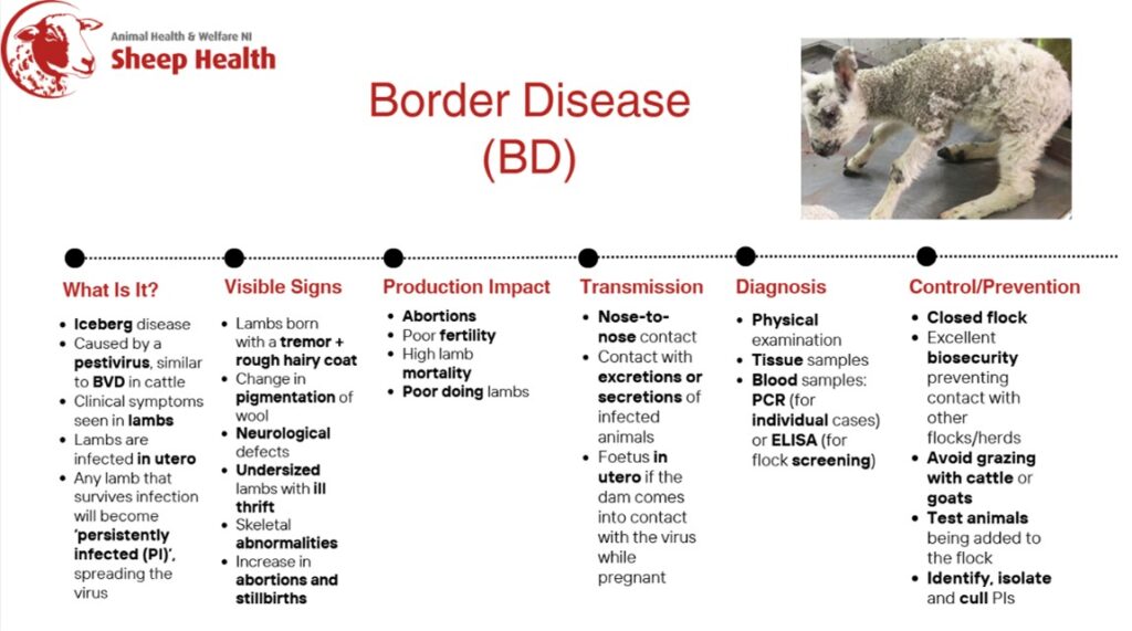
Border Disease (BD) is often referred to as ‘hairy shaker disease’. It’s caused by a pestivirus which is similar to the virus that causes BVD in cattle. It is possible that cross-infection can occur between cattle and sheep. Infection is most often seen causing infertility, abortion, foetal mummification, stillbirth and ill thrift lambs that sometimes display neurological signs. There is no treatment available. Infection results from in utero infection. When the virus is acquired in early pregnancy it infects the foetus by crossing over the placenta. If the foetus survives initial infection, the virus will infect foetal tissues causing a wide range of defects. Sometimes affected lambs can be born prematurely with many dying during the first weeks of life.
Any lambs that survive infection are known as ‘persistently infected.’ These animals will carry the virus their whole life and spread it to other animals. Sometimes, these animals can be born and show no symptoms, despite having the virus.
Infection has been estimated to be present in around a third of UK sheep flocks (FAS, 2020).
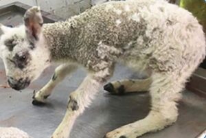
(Image credit: FAS, 2020)
Signs You May See in Your Flock
- Lambs born with a tremor (more pronounced during movement) and a rough, hairy coat
- Increase in crimp or change in pigmentation of wool
- High abortion levels and stillbirths
- Lambs with mild neurological defects
- Undersized lambs
- Skeletal abnormalities
- Ill thrift in lambs
Economic Losses
- Abortions
- Increased number of barren ewes
- High mortality in lambs
- Poor doing lambs
How Infection Spreads
- Direct contact- nose-to-nose contact with infected animals, saliva, urine, faeces, semen and blood
- If a pregnant ewe comes into contact with the virus, it will cross the placenta to infect the foetus, persistently infecting the lamb if it survives. If not born with the virus the pregnant ewe will not be infected for life.
Disease Diagnosis
Discuss physical symptoms with your private vet. Investigate any lamb that displays neurological signs such as shaking, unable or difficulty in standing, standing with a wide and unsteady stance or that are unusually hairy.
Tissue samples can be used from aborted or dead lambs, which will accurately confirm infection.
Blood testing using PCR (detects viral RNA) or viral antigen testing has a high sensitivity and specificity and, useful for diagnosis of individually infected lambs. Blood testing using antibody ELISA is useful for screening for evidence of circulating infection in adult sheep. This test is not useful in clinically affected lambs as PI animals are usually born without antibodies.
Prevention
- Keep a closed flock
- Biosecurity- avoid nose-to-nose contact with neighbouring flocks or herds
- Test all animals before adding them to the flock
- Keep cattle, goats and sheep separate, particularly during early pregnancy in ewes
Control
Identification, isolation and removal of PI animals
If BD is suspected in any lambs, they and their dam should be isolated immediately, ideally indoors. Both the dam and lamb should be tested. Any identified PIs must be culled.
Test for BD in all abortions and stillbirths
Abortions can be due to different diseases; therefore BD may go undetected for a period of time if other diseases have been responsible for abortions in the past.
Thorough disinfection
Disinfect all equipment used for the PIs.
Keep a closed flock and/or good biosecurity
Ensure that fencing and/or hedges are adequate so that neighbouring flocks cannot make direct nose-to-nose contact with your flock. Avoid grazing sheep with cattle or goats.
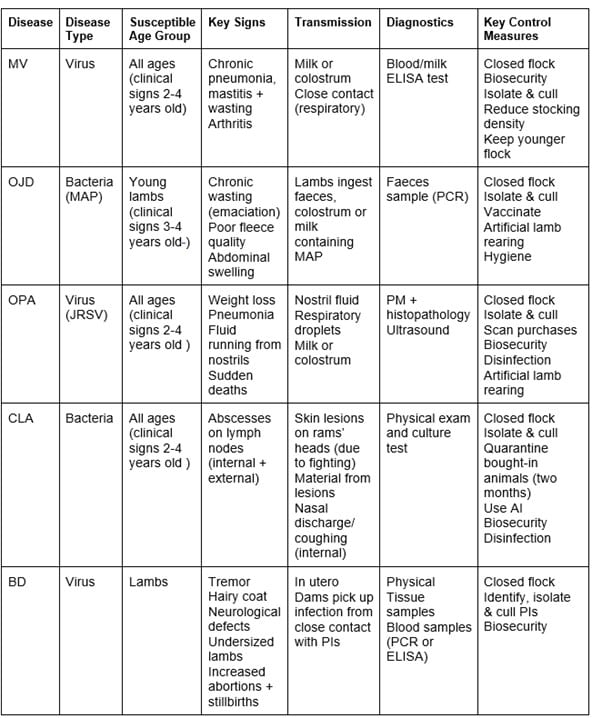
Iceberg Diseases Comparison
Decision Making Process- Do You Need To Test for Iceberg Diseases?